Additional Information
- We use a combination of images, videos, text and most importantly full dicoms where you can see and assess a scan just like you do at work.
- You can ask questions if you are unsure or need clarification at any stage of the course and Dr Ravi will reply to you. You can also see all the previous questions and answers asked by other registrants.
- There are multiple quizzes and review cases throughout the Mini Fellowship that will help you assess your understanding and retention of the learning.
- We make sure you understand the detailed and complex radiological anatomy of the Ankle, essential to an accurate diagnosis. We also cover the macroscopic pathology of abnormalities in all our Mini Fellowships, which most courses don't, but it makes understanding and reporting the imaging easier.
- There is also a pre workshop sequence of videos that will help you to learn and retain more.
- The focus of this course, like all our courses, is for you to be MORE CONFIDENT with your diagnosis, issue Clear Reports and to have your reports respected by the referring physician.
Accreditation: 30 CPD Hours by RANZCR
About Speaker
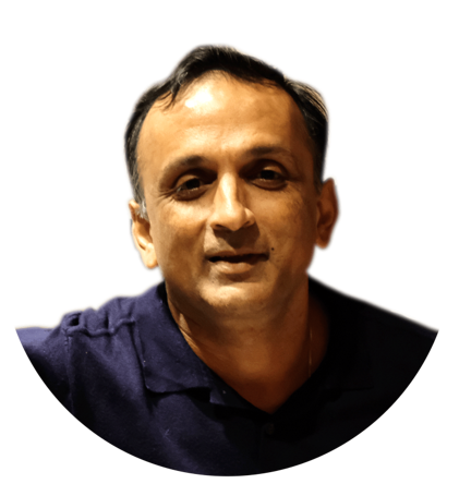
- Dr Ravi Padmanabhan
Dr Ravi Padmanabhan is the Director of Radiology Education Asia, and a Senior Consultant Radiologist from Australia, and now in Singapore. He has been teaching MSK and Spine MRI for over 10 years. His aim in the course is not just accumulating facts, but for you to be reporting more confidently at work.
His method of teaching is to simplify, without losing the essential things we need to know. For you to easily recognise the important anatomy, the relevant macroscopic pathology which helps to understand the imaging findings and for you to know where to look and what to look for. All of these help you to report a scan with confidence and issue reports that you are proud of and will be respected by referrers.